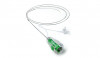
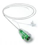
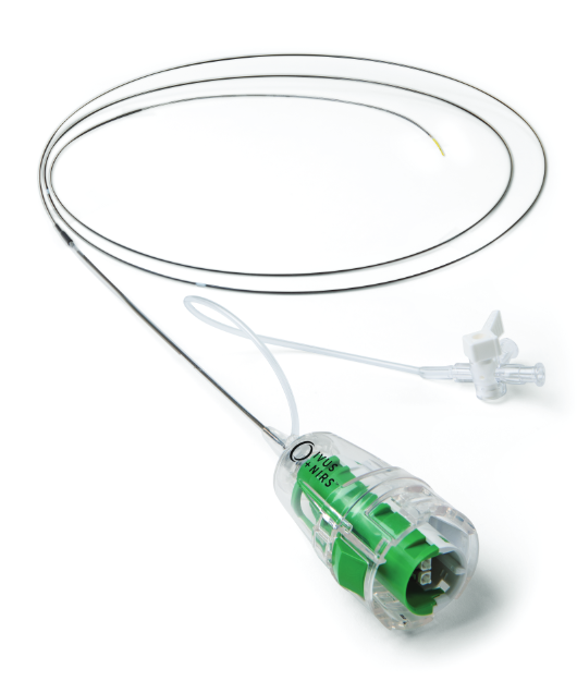

Together with the Makoto™ Intravascular Imaging System Dualpro™ IVUS+NIRS Catheter, is the only imaging system on the market today that utilizes not just intravascular ultrasound (IVUS) but also Near-Infrared Spectroscopy (NIRS) to help clinicians visualize vessel structure and gain valuable insights into plaque composition.
Specifications
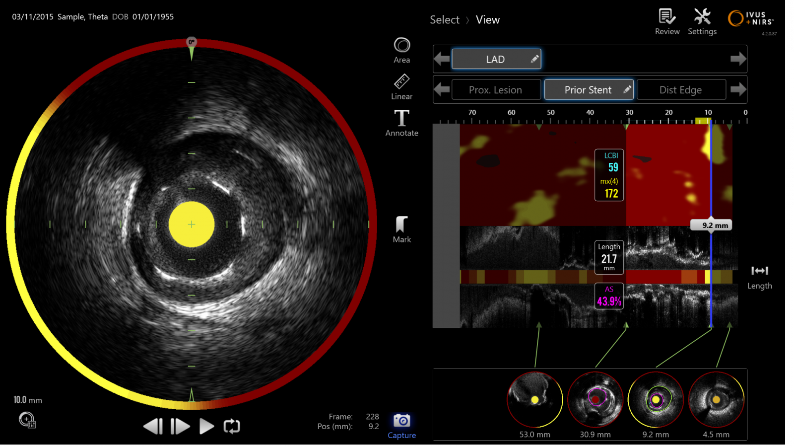
Make confident intervention decisions with the aid of the Lipid Core Burden Index (LCBI) and the simultaneous co-registered acquisition of IVUS and NIRS.
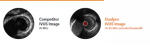
See a Crisper Image of Vessel Structure with the Dualpro IVUS

Dualpro™ is the only intravascular imaging catheter on the market today that utilizes Extended Bandwidth IVUS technology. By emitting and carefully processing a broad band of frequencies, the Dualpro™ IVUS brings you a best-in-class image resolution without compromising on the depth of field.
With a crisper IVUS image of the vessel structure, you can identify the degree of stenosis much more easily, visualize and quantify plaque burden, determine the landing zone for a stent, and assure proper stent deployment.
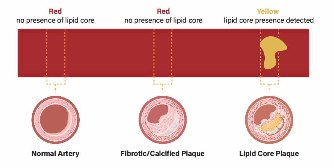
Dualpro™ is the only intravascular imaging catheter equipped with near-infrared spectroscopy (NIRS) technology. Now, you can easily identify unstable lipid core plaque (LCP) - a well-documented culprit in heart disease associated with 95% of STEM ls[MDL1] [AN2] and an increased risk of peri-procedural complications.
Why NIR Spectroscopy?
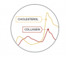
To identify lipids such as cholesterol, NIRS allows us to distinguish molecules, such as collagen and cholesterol, within the vessel wall and thus identify the presence of a lipid core plaque (LCP).
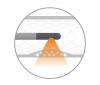
Where does the Light Propagate?
Through blood, tissue, and interstitial spaces. The microscopic mirrors at the tip of the Dualpro™ catheter are designed to deliver near-infrared light to the vessel wall and collect the diffusely reflected light.

How are the Spectra Interpreted?
Advanced algorithms analyze the returned light and calculate the probability of the presence of a lipid core plaque (LCP). Our algorithms have been validated in a large prospective histology study providing you with information you can trust.
IVUS image
References
- Detection by NIRCS or LCPS at culprit sites in Patients with Acute STEMI, Madder et al., JAC cardiovasc Interv. 2013
- Detection of LCPS by intracoronary NIRS identifies High Risk of Periprocedural MI, Goldstein et al., Circ cardiovasc Interv. 2011


Need help?
Get in touch
For information regarding Nipro products, services, and resource material:
| E-mail us [email protected] |
|
Submit a detailed inquiry: |
View our manufacturing locations: |